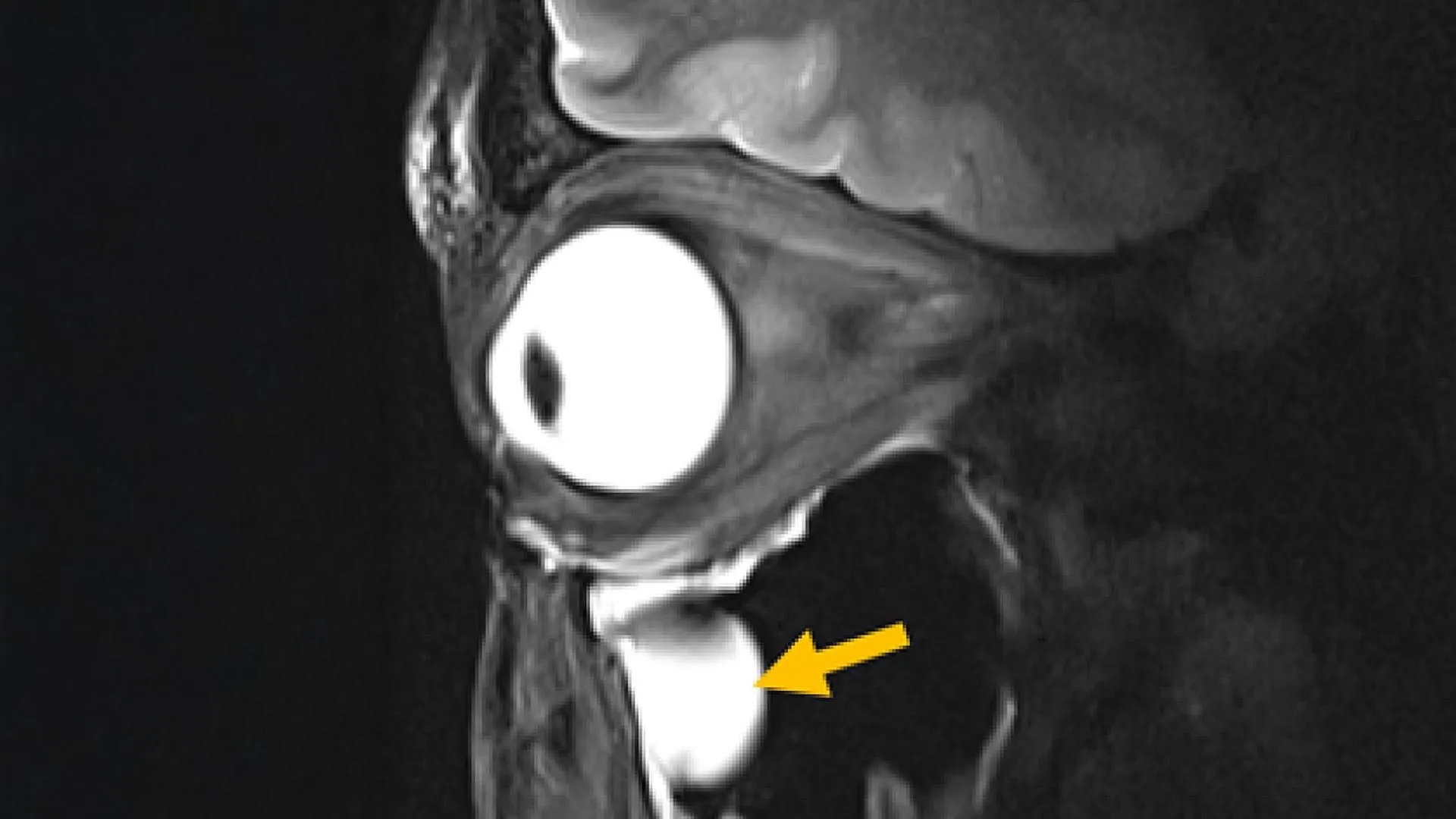
Doctors may soon get a clearer look inside the brain and eye thanks to a new MRI antenna that produces sharper images in less time. Researchers in Germany have developed the device using advanced engineered materials called metamaterials, which boost the radiofrequency signals that create MRI scans. The innovation, described in the journal Advanced Materials, could make scans faster and more detailed for patients with conditions affecting deep brain structures or delicate eye tissues.
MRI scanners work by sending radiofrequency signals into the body while a strong magnetic field is applied. Tissues respond to those signals, and the scanner collects the data to form an image. But traditional antennas, known as RF coils, often struggle to pick up enough signal from deep or anatomically complex areas, leading to blurry images and longer scan times. The new antenna incorporates metamaterials, which are specially engineered to interact with electromagnetic waves in ways natural materials cannot. In tests, the design strengthened signals from targeted tissues, increased spatial resolution, improved image sharpness, and accelerated data collection. The researchers tested the antenna on volunteers using a 7.0 Tesla MRI scanner to image the eye and orbit.
Because the antenna is compact and lightweight, it can be customized for different parts of the body and integrated into existing MRI systems without requiring expensive new machines. The team, led by Nandita Saha and Professor Thoralf Niendorf at the Max Delbrück Center, worked with experts from Rostock University Medical Center to validate the technology. “Our research demonstrates clear relevance for ophthalmological applications as it can facilitate anatomically detailed, high-spatial resolution MRI of the eye,” said Professor Oliver Stachs, a co-author at University Medicine Rostock. The technology may also help reduce unwanted heating around medical implants during scans and could improve MRI guided cancer treatments by directing radiofrequency energy more precisely for procedures such as tumor hyperthermia.
The research team is now preparing larger clinical studies involving multiple hospitals while modifying the antenna for additional organs, including the heart and kidneys. They also plan to adapt the design for MRI systems operating at different magnetic field strengths and for imaging atoms other than hydrogen, such as sodium and fluorine. “Innovations in imaging hardware have the potential to transform diagnostics, and this study is an important step toward next-generation MRI technology,” said Dr. Ebba Beller, a co-author at Rostock University Medical Center. With shorter scan times and sharper images on the horizon, patients could soon experience more comfortable exams and more confident diagnoses.